Abdominal Anatomy Female Left Side : Abdomen - Wikipedia : The muscle fibres of the diaphragm combine to form a central tendon.
byAdmin•
0
Abdominal Anatomy Female Left Side : Abdomen - Wikipedia : The muscle fibres of the diaphragm combine to form a central tendon.. The female reproductive system contains two main parts: This muscle is the innermost abdominal muscle. Jul 27, 2021 · distinguishing right from left is equally easy by using the liver as reference. Pregnant female with uterus displayed. Mar 05, 2012 · on the left side, the retrorenal plane and the perirenal space extend to the left diaphragm.
This muscle's origin is the lumbodorsal fascia and ribs. Feb 19, 2021 · abdominal pain is felt in the abdomen. Lower part of female urogenital tract. Welcome to the whitman college biology department's virtual pig dissection (vpd)! The hilum leads to a large cavity, called the renal sinus, within the kidney.
Pain Locator: Where Does it Hurt? from www.ligastrohealth.com Jul 27, 2021 · distinguishing right from left is equally easy by using the liver as reference. Welcome to the whitman college biology department's virtual pig dissection (vpd)! As was mentioned earlier, fluid collections in the paracolic and left inframesocolic spaces may communicate with the pelvic spaces ( fig 10 ). Lower part of female urogenital tract. The female reproductive system contains two main parts: This site is designed as a supplement to laboratory dissections exploring introductory mammalian anatomy and physiology — it is basic and many details have been omitted for clarity. Pregnant female with uterus displayed. This tendon ascends to fuse with the inferior surface of the fibrous pericardium.
It also laterally flexes and rotates the vertebral column.
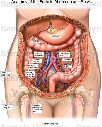
Pregnant female with uterus in situ. And the ovaries, which produce the anatomically female egg cells. The female reproductive system contains two main parts: As you know, this large organ is located on the right hand side of the abdomen, hence the left of the image is the patient's lateral right. This tendon ascends to fuse with the inferior surface of the fibrous pericardium. Pregnant female with uterus displayed. The ureter and renal vein leave the kidney, and the renal artery enters the kidney at the hilum. Lower part of female urogenital tract. Clinically the term abdominal cavity stands for abdominal cavity proper and thus we have separated abdomen and pelvis altogether for better understanding. As was mentioned earlier, fluid collections in the paracolic and left inframesocolic spaces may communicate with the pelvic spaces ( fig 10 ). This muscle is the innermost abdominal muscle. This muscle's origin is the lumbodorsal fascia and ribs. Mar 05, 2012 · on the left side, the retrorenal plane and the perirenal space extend to the left diaphragm.
The female reproductive system contains two main parts: The abdomen is an anatomical area that is bounded by the lower margin of the ribs and diaphragm above, the pelvic bone (pubic ramus) below, and the flanks on each side. This muscle is the innermost abdominal muscle. Either side of the pericardium, the diaphragm ascends to form left and right domes. Lower part of female urogenital tract.
Anatomy Of The Female Abdomen And Pelvis, Cut away View ... from healthiack.com Pregnant female with uterus displayed. The abdomen is an anatomical area that is bounded by the lower margin of the ribs and diaphragm above, the pelvic bone (pubic ramus) below, and the flanks on each side. Right horn dissected to show foetuses. As was mentioned earlier, fluid collections in the paracolic and left inframesocolic spaces may communicate with the pelvic spaces ( fig 10 ). The hilum leads to a large cavity, called the renal sinus, within the kidney. The abdominal cavity is subdivided by the plane of the pelvic inlet into a larger upper part, i.e., the abdominal cavity proper, and a smaller lower part, i.e., the pelvic cavity. And the ovaries, which produce the anatomically female egg cells. This site is designed as a supplement to laboratory dissections exploring introductory mammalian anatomy and physiology — it is basic and many details have been omitted for clarity.
As was mentioned earlier, fluid collections in the paracolic and left inframesocolic spaces may communicate with the pelvic spaces ( fig 10 ).
This muscle is the innermost abdominal muscle. Clinically the term abdominal cavity stands for abdominal cavity proper and thus we have separated abdomen and pelvis altogether for better understanding. The muscle fibres of the diaphragm combine to form a central tendon. In the adult, each kidney is approximately 3 cm thick, 6 cm wide, and 12 cm long. The abdominal cavity is subdivided by the plane of the pelvic inlet into a larger upper part, i.e., the abdominal cavity proper, and a smaller lower part, i.e., the pelvic cavity. Pregnant female with uterus in situ. Welcome to the whitman college biology department's virtual pig dissection (vpd)! It also laterally flexes and rotates the vertebral column. Jul 27, 2021 · distinguishing right from left is equally easy by using the liver as reference. As you know, this large organ is located on the right hand side of the abdomen, hence the left of the image is the patient's lateral right. And the ovaries, which produce the anatomically female egg cells. As was mentioned earlier, fluid collections in the paracolic and left inframesocolic spaces may communicate with the pelvic spaces ( fig 10 ). The hilum leads to a large cavity, called the renal sinus, within the kidney.
This muscle's origin is the lumbodorsal fascia and ribs. The ureter and renal vein leave the kidney, and the renal artery enters the kidney at the hilum. In the adult, each kidney is approximately 3 cm thick, 6 cm wide, and 12 cm long. It also laterally flexes and rotates the vertebral column. Either side of the pericardium, the diaphragm ascends to form left and right domes.
Anatomy M1 > Segal > Flashcards > Unit 25: Kidneys ... from classconnection.s3.amazonaws.com As was mentioned earlier, fluid collections in the paracolic and left inframesocolic spaces may communicate with the pelvic spaces ( fig 10 ). The abdomen is an anatomical area that is bounded by the lower margin of the ribs and diaphragm above, the pelvic bone (pubic ramus) below, and the flanks on each side. Either side of the pericardium, the diaphragm ascends to form left and right domes. The female reproductive system contains two main parts: This site is designed as a supplement to laboratory dissections exploring introductory mammalian anatomy and physiology — it is basic and many details have been omitted for clarity. Its insertion is at the pubis and linea alba (via aponeurosis), and its action is the compression of abdominal contents. Lower part of female urogenital tract. This tendon ascends to fuse with the inferior surface of the fibrous pericardium.
The female reproductive system contains two main parts:
Pregnant female with uterus displayed. It also laterally flexes and rotates the vertebral column. The abdominal cavity is subdivided by the plane of the pelvic inlet into a larger upper part, i.e., the abdominal cavity proper, and a smaller lower part, i.e., the pelvic cavity. The abdomen is an anatomical area that is bounded by the lower margin of the ribs and diaphragm above, the pelvic bone (pubic ramus) below, and the flanks on each side. The muscle fibres of the diaphragm combine to form a central tendon. The hilum leads to a large cavity, called the renal sinus, within the kidney. Pregnant female with uterus in situ. Right horn dissected to show foetuses. As you know, this large organ is located on the right hand side of the abdomen, hence the left of the image is the patient's lateral right. This muscle's origin is the lumbodorsal fascia and ribs. The ureter and renal vein leave the kidney, and the renal artery enters the kidney at the hilum. The female reproductive system contains two main parts: This muscle is the innermost abdominal muscle.
The abdomen is an anatomical area that is bounded by the lower margin of the ribs and diaphragm above, the pelvic bone (pubic ramus) below, and the flanks on each side abdominal anatomy. Clinically the term abdominal cavity stands for abdominal cavity proper and thus we have separated abdomen and pelvis altogether for better understanding.